Trisoma®
CranioSacral Therapy
When people think of circulation, the cardiovascular (blood) system usually comes to mind. However humans also have three other important circulatory systems: lymphatic, glymphatic and craniosacral.
Craniosacral Therapy is a type of bodywork that uses subtle palpation and intuitive "listening" to detect the movement and restrictions in the cerebrospinal fluid and various related structures to facilitate well-being. It is typically an excellent modality for clients who are limited by medications or physical conditions from receiving deep manual tissue bodywork. Clients remain clothed, and no oil is used.

The craniosacral (or cranial-sacral) system is composed of the, cranium (skull), meninges, vertebrae (spine), cerebrospinal fluid and all the structures related to the production, reabsorption and containment of cerebrospinal fluid. According to the Ohio State University Medical Center[1], the craniosacral system has a rhythmic motion, normally between 6 to 12 cycles per minute, which helps propel cerebral spinal fluid throughout the central nervous system between the center of the brain, around the brain, down the spine to the sacrum. The 20 plus bones that comprise the skull and face fit together in such a way as to allow minute movement throughout life. This movement and relationship between the bones of the skull are essential to maintaining the normal pumping action of cerebral spinal fluid.
A normally functioning craniosacral system is essential to a healthy central nervous system. Because the nervous system affects all other body systems, manipulation of the craniosacral system can influence all parts of the body. Budiman Minasny, PhD of the University of Sydney, Australia, proposes a hypothetical model that builds on the neurobiologic, ideomotor action, and consciousness theories, where the therapist stimulates mechanoreceptors in the fascia, resulting in ideomotor action and fascial or myofascial unwinding. [11]. The Cleveland Clinic calls this CST tension relief "fascial clearance" [10]. More on the developing research on fascia is at our Myofascial Release page. .

About a pint (~500 ml) of cerebrospinal fluid (CSF) is produced
...every day in the choroid plexus of the brain, and it circulates through the brain, through the spinal cord to the sacrum. Cerebrospinal fluid has many putative roles including mechanical protection of the brain, facilitation of pulsatile cerebral blood flow, distribution of peptides, hormones, neuroendocrine factors (now implicated in obesity) and other nutrients and essentials substances to cells of the body, and in turn wash away waste products. Cardiovascular dynamics are also affected by CSF pressure, as the flow of blood must be tightly regulated within the brain to assure consistent brain oxygenation. CSF has a pulse, or variance in rate of flow, of its own, distinct from that of the heartbeat or the breath.
The CSF pulse
...is said to be felt anywhere on the body by a sensitive practitioner, but most palpation is done on the head and sacrum. The touch used is very light and gentle, but because we are working with the fluid that facilitates constant nourishment, including chemicals which promote Alzheimer's Disease, and multi-modal communication within the body, Craniosacral Therapy may yield profound unwinding as if unraveling a tangled knot. [9] This unwinding and releasing may be felt as a relief on both physical and emotional levels. Some postulate that a human Life Force (a.k.a. breath of life, or Qi) is a main fixture in healing and is transported via the cerebrospinal fluid (CSF) to all the tissues of the body. Thus, this fluid is involved in the healing of all of the major organs and organ systems of the body.
Disorders of CSF flow
...may therefore impact not only CSF movement, but also the intracranial blood flow, with subsequent neuronal and glial vulnerabilities. The venous system is also important in this equation. Infants and patients shunted as small children may have particularly unexpected relationships between pressure and ventricular size, possibly due in part to venous pressure dynamics. This may have significant treatment implications but the underlying pathophysiology needs to be further explored.

CSF connections with the lymphatic system
... have been demonstrated in several mammalian systems. Preliminary data suggest that these CSF-lymph connections form around the time that the CSF secretory capacity of the choroid plexus is developing (in utero). There may be some relationship between CSF disorders, including hydrocephalus and impaired CSF lymphatic transport. And of course the recently discovered glymphatic system is attracting much research.
History
Andrew Taylor Still, born in Virginia in 1828, engineer and army surgeon during the American Civil War, became dispirited with the medical shortcomings of his day, and is credited with promoting what is now called Osteopathy. In the 1930's William Sutherland, American osteopath, was credited as first researching Cranial Osteopathy and Primary Respiratory Impulse (PRI) , and discovered that the cerebrospinal fluid surrounding the brain and spinal chord flowed in a rhythmic cycle. In the last century, John E. Upledger coined the term Craniosacral Therapy, and he, Milne and others theorized that adult cranial bones do, in fact, move slightly at the sutures. More recent evidence supports such theory of cranial bone mobility.
These osteopaths developed refined techniques for very subtle and gentle manual facilitation of the skull and sacrum which have been shown to affect structural positions that affect these fluid fluctuations. I studied with Paul Brown of the Milne Institute, which is continuing to develop this therapy. This modality is now accepted by health insurance in several countries in Europe for various idiopathic and other conditions, and can be incorporated with other modalities which may be covered by US insurance..
Unsettled Science
CST continues to be controversial due to limited research studies, but it is practiced in US hospitals and medical centers, such as Wexner Medical at Ohio State University. In the 2000's when I was in cadaver class, the anatomy professor still taught that the vermiform appendix is a useless vestigial organ, although now many agree that it is a hiding place for probiotics and infection-fighting lymphoid cells. Newer theories state that it lubricates the ileocecal valve, perhaps as an enigmatic part of the ileocecal region and even Leonardo Da Vinci described its function as a relief valve.
Similarly, it has been taught in medical schools that cranial bones fuse by adulthood, however recently it has been shown that the cranial bones in a living human do not completely fuse, but allow minute movement. In 2002 at the Philadelphia College of Osteopathic Manipulative Medicine, research measured cranial bone mobility by x-ray. [7]

Recent research
... indicates that the nervous system, fascia, lymph and glymph communicate in other ways than just nerve impulses. It is theorized that environmental factors, such as electromagnetic fields, fluorescent lights, cell phones, ultrasound and stress may be seriously affecting our nervous system health. In fact, US Patents have been granted for controlling brain activity with microwaves and ultrasound. When trauma or disease prevent someone from receiving other types of bodywork, this modality may be ideal to try. Although Paul has a technical background, he does not claim to know what happens in a particular C-S Therapy session; no quantum mechanics or tachyons are claimed, as some other practitioners have stated.
U.S. National Library of Medicine and the National Institutes of Health published a 2004 study that tested orthopedic versus craniosacral treatment for tennis elbow. In this study it was possible to successfully treat the chronic Epicondylopathia humeri radialis with an osteopathic approach. A significant difference to an orthopedic treatment could not be proved. [3]
Although medical schools have long taught that the cranial sutures are fixed after childhood, Cranio: the journal of craniomandibular practice, reported in 1992 that: "Craniosacral therapy supports that light forces applied to the skull may be transmitted to the dura membrane having a therapeutic effect to the cranial system. This study examines the changes in elongation of falx cerebri during the application of some of the craniosacral therapy techniques to the skull of an embalmed cadaver. The study demonstrates that the relative elongation of the falx cerebri changes as follows: for the frontal lift, 1.44 mm; for the parietal lift, 1.08 mm; for the sphenobasilar compression, -0.33 mm; for the sphenobasilar decompression, 0.28 mm; and for the ear pull, inconclusive results. The present study offers validation for the scientific basis of craniosacral therapy and the contention for cranial suture mobility."
The Journal of Neuropathology and Applied Neurobiology reported in 2003 that "There is mounting evidence that a significant portion of cerebrospinal fluid drainage is associated with transport along cranial and spinal nerves with absorption taking place into lymphatic vessels external to the central nervous system... linking the subarachnoid compartment with extracranial lymphatics. [4]
The glymphatic system is being fervently researched in relation to fluid flow in the human brain.
German neurologists showed evidence for inspiration as the major regulator of human CSF flow. [6]
Additionally it should be noted that there was a study refuting the effects of cranial manipulation on CSF pressure and movement between sutures, however this study involved machinery compressing the skulls of anesthetized rabbits. [5]
Quotes
"Because we don't understand the brain very well we're constantly tempted to use the latest technology as a model for trying to understand it. In my childhood we were always assured that the brain was a telephone switchboard. (What else could it be?) And I was amused to see that Sherrington, the great British neuroscientist, thought that the brain worked like a telegraph system. Freud often compared the brain to hydraulic and electromagnetic systems. Leibniz compared it to a mill, and now, obviously, the metaphor is the digital computer." -John R. Searle, philosophy professor (1932- )
References
1.
Ohio State University Medical Center website, Services / Procedures, CranioSacral Therapy,
Address
2000 Kenny Road
Columbus, OH 43221.
Retrieved Jan. 2008, from http://medicalcenter.osu.edu/patientcare/healthcare_services/services/?ID=1491
(Return to Reference 1 in text)
2.
Cranio. 1992 Jan;10(1):9-12.Links
Changes in elongation of falx cerebri during craniosacral therapy techniques applied on the skull of an embalmed cadaver.
Kostopoulos DC, Keramidas G.
PMID: 1302656 [PubMed - indexed for MEDLINE]
3.
Geldschläger S.
Forsch Komplementarmed Klass Naturheilkd. 2004 Apr;11(2):93-7.
Abstract
[Osteopathic versus orthopedic treatments for chronic epicondylopathia humeri radialis: a randomized controlled trial]
Forsch Komplementarmed Klass Naturheilkd. 2004 Apr;11(2):93-7. German.
www.ncbi.nlm.nih.gov
PMID: 15138373 [PubMed - indexed for MEDLINE]
4. Zakharov A, Papaiconomou C, Djenic J, Midha R, Johnston M (2003).
"Lymphatic cerebrospinal fluid absorption pathways in neonatal sheep revealed by subarachnoid injection of Microfil".
Neuropathol. Appl. Neurobiol. 29 (6): 563-73.
5.
J Orthop Sports Phys Ther. 2006 Nov;36(11):845-53.Links
Craniosacral therapy: the effects of cranial manipulation on intracranial pressure and cranial bone movement.
Downey PA, Barbano T, Kapur-Wadhwa R, Sciote JJ, Siegel MI, Mooney MP.
Physical Therapy Program, Chatham College, Woodland Rd, Pittsburgh, PA 15232, USA. downey@chatham.edu
PMID: 17154138 [PubMed - indexed for MEDLINE]
6.
Cranio 1992 Jan;10(1):9-12. doi: 10.1080/08869634.1992.11677885.
D C Kostopoulos, G Keramidas
Changes in elongation of falx cerebri during craniosacral therapy techniques applied on the skull of an embalmed cadaver
From https://pubmed.ncbi.nlm.nih.gov/1302656/
(Return to Reference 6 in text)
7.
Sheryl Lynn Oleski , Gerald H. Smith & William T. Crow (2002)
Radiographic Evidence of Cranial Bone Mobility, CRANIO®,
The Journal of Craniomandibular & Sleep Practice
Volume 20, 2002 - Issue 1,
Philadelphia College of Osteopathic Manipulative Medicine, PA 19131, USA.
20:1, 34-38, DOI: 10.1080/08869634.2002.11746188
(Return to Reference 7 in text)
8.
Hodge BD, Kashyap S, Khorasani-Zadeh A.
Anatomy, Abdomen and Pelvis: Appendix. [Updated 2023 Aug 8].
In: StatPearls [Internet]. Treasure Island (FL): StatPearls Publishing; 2024 Jan-.
Available from: https://www.ncbi.nlm.nih.gov/books/NBK459205/
(Return to Reference 8 in text)
9.
Anecdotal description from client, 2008.
(Return to Reference 9 in text)
10.
Craniosacral Therapy page.
Cleveland Clinic, the world’s first integrated international health system.
With more than 65,000 caregivers worldwide, Cleveland Clinic has almost 6 million patient visits per year, at more than 200 locations.
Available 04/30/2024 from: https://my.clevelandclinic.org/health/treatments/17677-craniosacral-therapy
(Return to Reference 10 in text)
11.
Minasny B. Understanding the process of fascial unwinding.
Int J Ther Massage Bodywork. 2009 Sep 23;2(3):10-7.
doi: 10.3822/ijtmb.v2i3.43. PMID: 21589734; PMCID: PMC3091471.
https://www.ncbi.nlm.nih.gov/pmc/articles/PMC3091471/
(Return to Reference 11 in text)
Images
1.
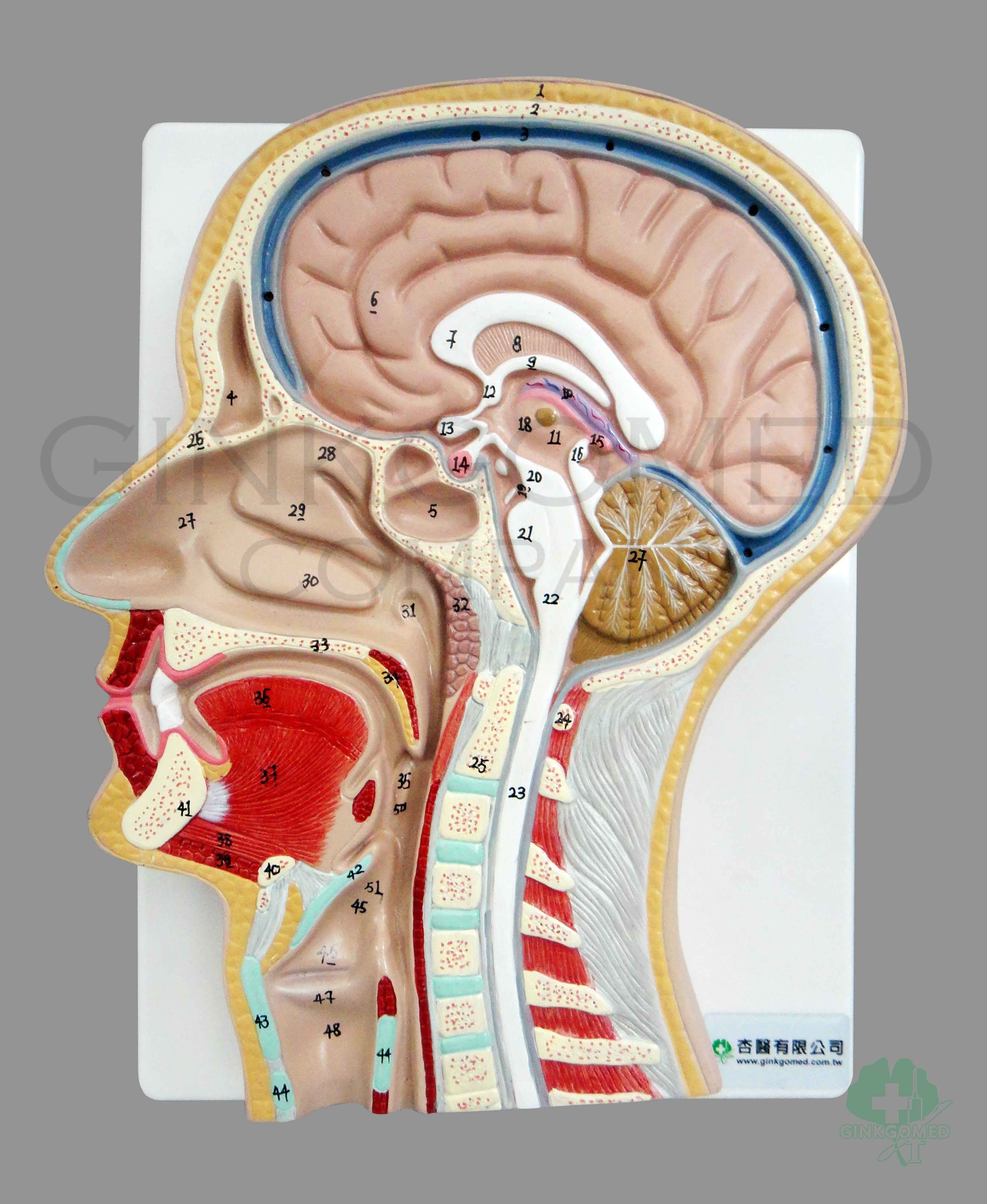
Image of PVC relief model of mid-sagittal section of head and neck by Ginkgomed Company (link to sales page)
(Return to Image 1 in text)
2.
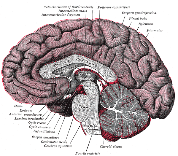
Image of the Fore-brain or Prosencephalon, by
Gray, Henry.
Anatomy of the Human Body.
Philadelphia: Lea & Febiger, 1918; Bartleby.com, 2000. www.bartleby.com/107/.
(Return to Image 2 in text)
3.
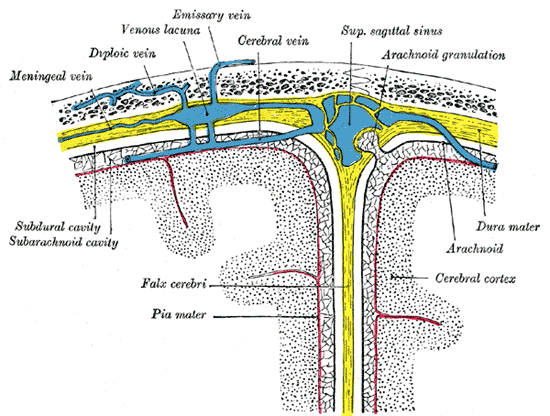
Image of Diagrammatic representation of a section across the top of the skull, showing the membranes of the brain, etc. (Modified from Testut.), by
Gray, Henry.
Anatomy of the Human Body. Philadelphia: Lea & Febiger, 1918; Bartleby.com, 2000. www.bartleby.com/107/.
(Return to Image 3 in text)
4.
Image of spine, by
Gray, Henry.
Anatomy of the Human Body. Philadelphia: Lea & Febiger, 1918; Bartleby.com, 2000. www.bartleby.com/.
(Return to Image 4 in text)
"If it works, do it. If you try to understand everything, you will make yourself crazy." - Dr. Jean-Louis Durant (French-American osteopath) to Paul on 2006 May 31.